

IV Cannula: What It Is, Types, Sizes & How It’s Inserted
This guide is for general education. It is not clinical instruction. Always follow the manufacturer’s instructions for use and your hospital’s protocol.
An IV cannula is one of the most frequently used devices in hospitals, emergency departments and operating theatres. Patients often only notice it as a small plastic hub taped to the hand, but it is what makes fast, reliable treatment possible. This guide covers what an IV cannula is, why and when it is used, the types and sizes available, and how it is placed.
What is an IV cannula?
An (intravenous) IV cannula is a small , flexible tube inserted into a peripheral vein , usually in the hand or forearm . Using a needle, the catheter is guided into the vein and then needle is withdrawn, leaving only the soft catheter in place. This allows clinicians to access the bloodstream directly for fluids, medicines, blood and blood products.
This process of putting IV Cannula in is called IV cannulation.
Why IV cannulation is done
- Speed. Medicines put into a vein go right into the blood stream. It counts in emergencies, intensive care and surgery.
- If oral treatment is not an option: oral drugs are not reliably absorbed in patients who cannot swallow, are vomiting, or are admitted in ICU’s.
- Continuous access: One cannula allows the medical team to administer multiple medicines over hours or days without the need for a new needle stick each time.
- Dose control Infusions allow the drug dose and the speed of the infusion to be set and adjusted accurately.
- Comfort: Less pain and vein irritation for patients requiring frequent therapy due to less repeated injections.
When is an IV cannula needed?
- Body Fluids: To replace fluid lost through dehydration, blood loss or shock, and to help maintain blood pressure and electrolyte balance.
- Medicines: to administer Antibiotics, pain relief, chemotherapy, sedatives and anaesthetic medicines directly into the bloodstream to control the situation of the patient.
- Emergencies. Trauma, severe infection, cardiac arrest or any situation needing immediate drug access.
- Blood transfusion: Used for anaemia, surgical blood loss or trauma.
- Children and Newborns: Infants and children need IV therapy for infection, hydration or nutrition and use smaller gauges.
High quality cannulas like the IV Cannula from GSTC are developed to assist in these cases and to reduce patient pain.
Parts of an IV cannula:
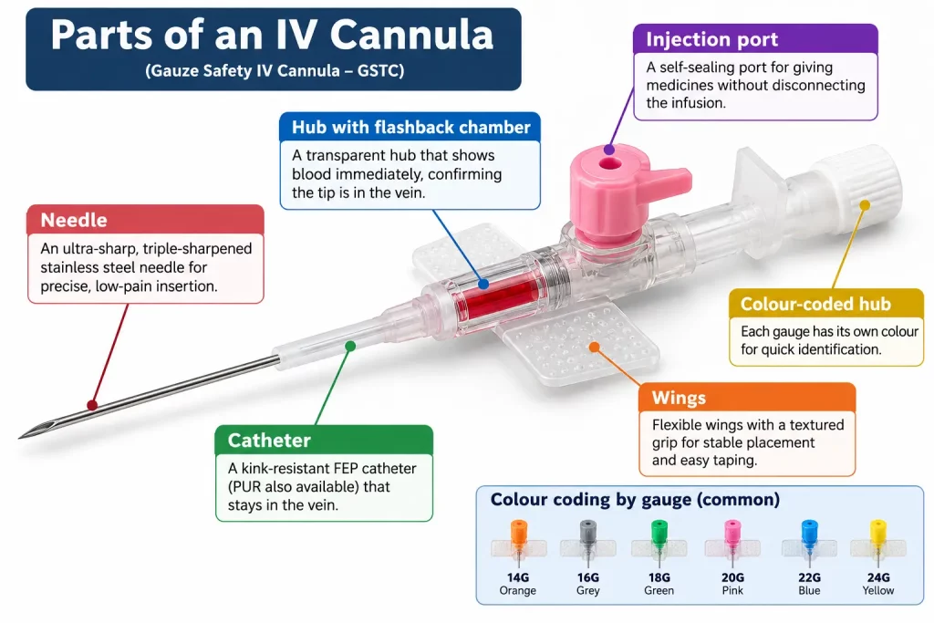
A typical GSTC IV cannula has
- Needle: Ultra-sharp, triple-sharpened stainless steel needle for precise, low-pain insertion.
- Catheter : Kink resistant FEP catheter (PUR also available) which stays in the vein.
- Hub with flashback chamber: Transparent hub which shows blood immediately confirming that the tip is in the vein.
- Wings: Textured grip for stable placement and easy taping flexible wings.
- Injection port: A port that seals itself and is used to give medicines without disconnecting the infusion.
- Colour coded hub: Each gauge is colour coded for easy identification.
 Types of IV cannula
- I.V. Cannula with wings and port. The most common all-purpose design. The wings aid in fixation and the port permits intermittent medication.
- I.V. Cannula with No wings, no port (pen type): simple, low profile, often used for short procedures and quick access.
- Safety I.V. Cannula: built-in needle shield minimises needlestick injuries to staff. Many hospitals prefer these now.
- Paediatric and neonatal cannulas: Smaller gauges, typically 22G to 26G, for small and fragile veins
Catheter material is also a factor. PUR softens at body temperature and can be more gentle on the vein. FEP is somewhat stiffer and resists kinking. The right choice depends on how long the device stays in and on the hospital protocol..
IV cannula sizes: gauge, colour and flow rate
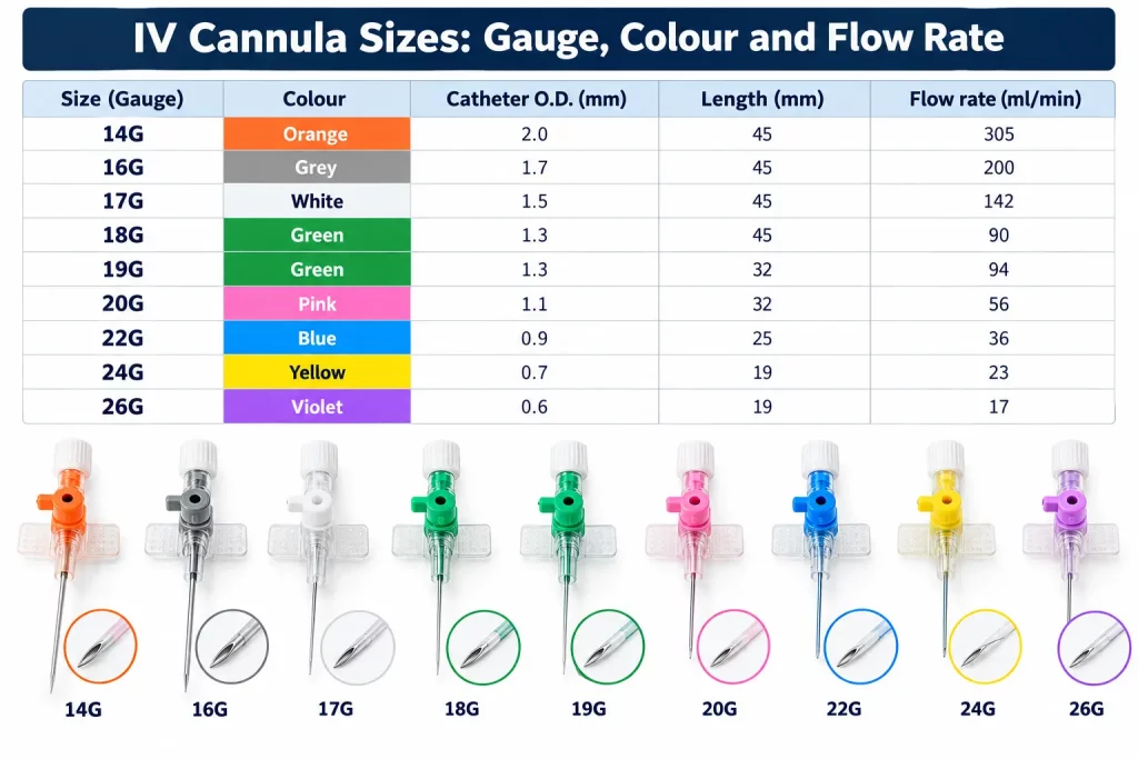
Size is given in gauge (G). A lower number means a wider catheter and a faster flow. GSTC’s range covers 14G to 26G.
| Size | Colour | Catheter O.D. | Length | Flow Rate (ml/min) |
|---|---|---|---|---|
| 14G | Orange | 2.0 mm | 45 mm | 305 |
| 16G | Grey | 1.7 mm | 45 mm | 200 |
| 17G | White | 1.5 mm | 45 mm | 142 |
| 18G | Green | 1.3 mm | 45 mm | 90 |
| 19G | Green | 1.3 mm | 32 mm | 94 |
| 20G | Pink | 1.1 mm | 32 mm | 56 |
| 22G | Blue | 0.9 mm | 25 mm | 36 |
| 24G | Yellow | 0.7 mm | 19 mm | 23 |
| 26G | Violet | 0.6 mm | 19 mm | 17 |
Choosing a gauge:
- 14G-16G: rapid fluid and blood transfusion
- 18G-20G: general adult IV access and surgery
- 22G-24G: paediatric and elderly patients
- 26G: neonates and fragile veins
When is an IV cannula used?
- IV cannula helps to cure dehydration & blood loss by providing direct access to the bloodstream.
- Administration of Antibiotics, pain relief and other drugs that need to be delivered quickly or reliably
- Transfusion of blood and blood components
- Perioperative Care and Anaesthesia
- For Emergency access in Trauma, Sepsis or Cardiac Arrest.
Role of Quality IV Cannulas in Patient Safety
- Smooth insertion
- Less trauma to veins
- Lower infection risk
- Better. patient experience
Not all IV cannulas are the same. Quality matters when it comes to GSTC IV Cannula, they are manufactured with high precision to ensure smooth cannulation and reliable flow rates, making it best suited for both emergency and routine use.
Choosing a site, and where not to place one
When it comes to access the vien for the transfusion or any other treatment, Medical professional usually start with the forearm or the back of the hand and prefer veins that are straight, soft and not over a joint. In infants, scalp or foot veins may be used.
Sites generally avoided include:
- An arm with a dialysis fistula or graft
- The side affected by lymphoedema or after axillary lymph node removal
- Areas that are bruised, inflamed, infected or hard
- The lower limbs in adults, because of higher infection and thrombophlebitis risk
How IV cannulation is done: an overview
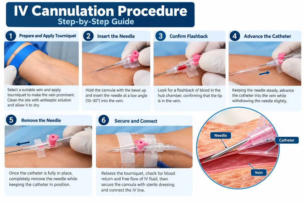
Cannulation must be performed by trained clinicians following local protocol. In outline:
- Prepare: in this medical professional prepares the patient, find the patient vein by tightening his or her hand and accessing the vein access.
- Tourniquet: Apply 7-10 cm above the site.
- Choose a vein: Straight, springy, well-filled (hand or forearm preferred). Avoid bruised, infected, or fistula/mastectomy-side limbs.
- Clean with alcohol or chlorhexidine for 30 seconds; let it dry. Don’t re-palpate.
- Glove up and anchor the skin taut below the site.
- Insert bevel up at 10-30° until you see flashback.
- Flatten the angle, advance 1-2 mm, then slide the catheter off the needle into the vein.
- Release the tourniquet, press lightly over the vein tip, and remove the needle into the sharps bin.
- Flush with saline. Stop if there’s pain, swelling, or resistance.
- Secure with a dressing.
Complications and how they are reduced
| Complication | What It Is | Main Prevention |
|---|---|---|
| Phlebitis | Inflammation and pain in the vein | Choose the correct gauge, use aseptic technique, and perform regular checks |
| Infiltration | IV fluid leaking into the surrounding tissue | Secure the cannula properly and remove promptly if swelling appears |
| Extravasation | Medication leaking into the surrounding tissue | Confirm correct placement and monitor the IV site closely |
| Haematoma | Bruising caused by vein puncture | Use a gentle insertion technique and apply pressure after removal |
| Infection | Local or bloodstream infection | Maintain hand hygiene, proper skin antisepsis, and dressing integrity |
Regular site assessment matters more than any single device feature. Redness, pain, swelling or leakage are all reasons to review the cannula.
How long can an IV cannula stay in?
The usual practice was to change peripheral cannulas every 72 to 96 hours. More recent evidence suggests replacement if clinically indicated . Hospital policies vary . Follow local protocol.
How GSTC manufactures IV cannulas
GSTC manufactures IV cannulas in 14G-26G under an ISO 13485:2016 quality system. Each cannula is sterile and individually packaged, and all batches are tested for sterility, leakage, flow rate and tip sharpness. For specifications and export enquiries, see our IV cannula product page.
References
- World Health Organization. Guidelines for the prevention of bloodstream infections and other infections associated with the use of intravascular catheters. Part 1: peripheral catheters. Geneva: WHO; 2024.
https://www.who.int/publications/i/item/9789240093829 - Nickel B, Gorski L, Kleidon T, et al. Infusion Therapy Standards of Practice, 9th Edition. Journal of Infusion Nursing. 2024;47(1S Suppl 1):S1-S285.
https://doi.org/10.1097/NAN.0000000000000532
PubMed: https://pubmed.ncbi.nlm.nih.gov/38211609/ - O’Grady NP, Alexander M, Burns LA, et al. Guidelines for the Prevention of Intravascular Catheter-related Infections. Clinical Infectious Diseases. 2011;52(9):e162-e193.
https://pmc.ncbi.nlm.nih.gov/articles/PMC3106269
CDC copy (last updated October 2017): https://stacks.cdc.gov/view/cdc/134991 - Webster J, Osborne S, Rickard CM, Marsh N. Clinically-indicated replacement versus routine replacement of peripheral venous catheters. Cochrane Database of Systematic Reviews. 2019;1:CD007798.
https://doi.org/10.1002/14651858.CD007798.pub5
PubMed: https://pubmed.ncbi.nlm.nih.gov/30671926/ - International Organization for Standardization. ISO 10555-5:2013 Intravascular catheters: Sterile and single-use catheters. Part 5: Over-needle peripheral catheters.
https://www.iso.org/standard/54885.html